
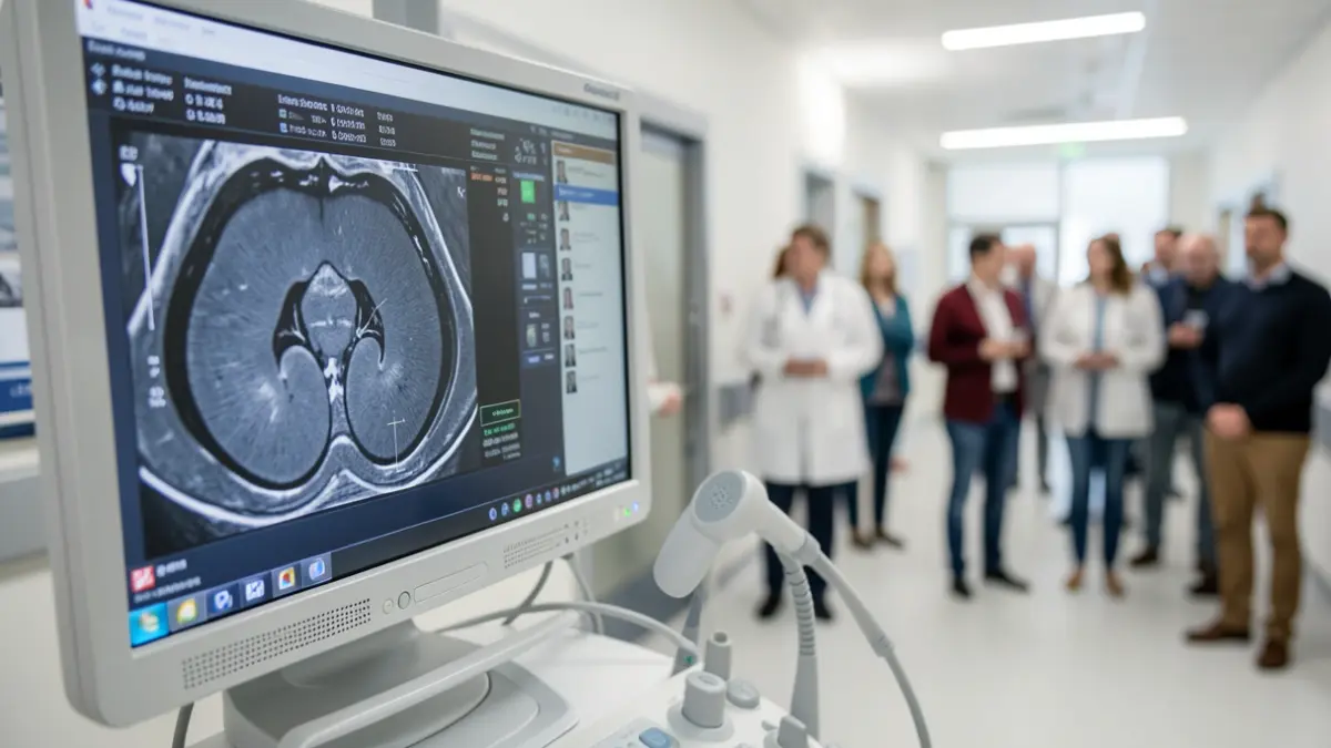
The technique allows for real-time combination of images obtained through magnetic resonance imaging with those from the ultrasound used during the biopsy. The aim is to improve diagnostic performance and reduce the possibility of false negatives in locating suspicious prostate areas.
Previously, when an MRI showed a suspicious lesion, the specialist would use the prior images as a reference to locate the area for biopsy using ultrasound, a procedure known as cognitive biopsy.
The new equipment incorporates specific software that imports MRI images and fuses them with the ultrasound in real-time. This allows the specialist to visualize the previously identified area and direct the sample collection towards it.
“"Instead of biopsying an area we believe is there, the overlay of the MRI and ultrasound image precisely indicates the area to be biopsied."
The technique will primarily be applied to patients whose prostate MRI shows a suspicious area. When the MRI does not identify a lesion suitable for targeted biopsy, the standard techniques will be maintained. Patients with suspicious findings will be included in a specific fusion biopsy schedule.
The Urology Department has begun integrating the technique into its care activities after performing initial procedures and plans to progressively increase its use based on clinical indications.